Nov 4, 2012
Atrial fibrillation
Atrial fibrillation can be either persistent or paroxysmal. When dealing with new onset atrial fibrillation (AF), generally we tend to wait for at least a week before labelling it as persistent AF. The treatment of persistent AF is either rate-control or rhythm-control. When we choose the rate-control strategy, we allow the arrhythmia to persist (atrial rate in AF is more than 350 per minute) but we use drugs to prevent the ventricular rate from exceeding a particular limit (below 90/min at rest and below 110/min during usual activity).
When we choose the rhythm-control strategy, we take measures to change the AF to sinus rhythm. We do this either with drugs (usually Class 1C or Class 3 antiarrhythmic drugs) or by electrical means. Research on atrial fibrillation has told us that both the rate-control and rhythm-control strategies are fine provided patients on rate-control are on adequate long term anticoagulation. It does seem a bit illogical that allowing a fast atrial arrhythmia to remain intact has the same outcomes as changing it back to its normal rhythm. The answer lies in knowing that long term use of antiarrhythmic drugs is not completely safe because they have a pro-arrhythmic effect and can provoke ventricular arrhythmias which lead to complications.
We also know that if we can control the ventricular rate adequately, we can prevent tachycardia-induced cardiomyopathy from developing.
For those who have paroxysmal AF, the treatment options are not so clearly defined even though we know that long term anticoagulation is necessary for them too. Patients with paroxysmal AF tend to be treated with antiarrhythmic drugs. Another option for such patients is catheter-ablation therapy, a procedure where the pulmonary veins in the left atrium are electrically isolated by using radio-frequency energy. All invasive procedures have risks and catheter ablation for AF can lead to strokes (by dislodging microemboli from the left atrium) and cardiac tamponade (by piercing the atrial wall).
A recent study in the New England Journal of Medicine tells us that both drug therapy and catheter ablation are effective for patients with paroxysmal AF and that there is no strong evidence yet that one is better than the other even though we know that those who undergo catheter ablation have less recurrence of AF after two years.

Oct 17, 2012
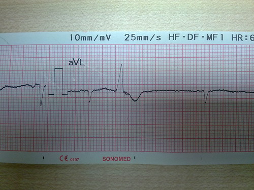
Question: Is it possible to say what the cardiac axis might be just by looking at Lead aVL as shown above?
Answer: It is not possible to say exactly what the cardiac axis is but one can infer in this manner:
1. The current is moving away from aVL. Left axis is the state where the current is moving towards the left side in the segment beyond minus 30 degrees. Therefore lead aVL tells me that this is not left axis.
2. If the current is exactly minus 30 degrees (normal axis) or zero degrees (normal axis), or plus 30 degrees (normal axis), lead aVL should be having a net positive deflection because the electrical scatter is still towards aVL. Therefore the axis is not zero degrees or minus 30 degrees or plus 30 degrees.
3. If the axis is plus 60 degrees, lead aVL should have biphasic deflection (positive and negative deflections are equal) because plus 60 degrees is exactly perpendicular to Lead aVL. Therefore the axis is not plus 60 degrees either.
4. If the axis is plus 90 degrees (normal axis) or beyond 90 degrees (right axis), lead aVL will show a negative deflection because now the electrical scatter is truly away from aVL. Therefore the axis here may be normal (between plus 60 and 90 degrees) or right axis (beyond plus 90 degrees).
Question: Is the ventricular ectopic seen in the given Lead aVL, arising from the right ventricle (RV) or from the left ventricle (LV)?
Answer: If an ectopic beat starts in the RV, it will depolarise the RV first and the LV later. Hence a ventricular ectopic that originates from the RV will have a LBBB pattern. Similarly, a left ventricular ectopic depolarises the LV first and the RV later. Hence it will show a RBBB pattern. So, if a ventricular ectopic shows the LBBB pattern, it is a ventricular ectopic from the RV and if the ectopic beat shows a RBBB pattern, it is a left ventricular ectopic.
The ectopic beat in aVL here (a lead that looks at the left side of the heart) has an LBBB pattern. Therefore it is an ectopic beat that originates from the RV.
Sep 11, 2012
A man who could not feel the left side of his body
A 50 year old man was admitted with a sudden inability to feel the left side of his face, left arm and left lower limb. He could speak and he could move all his limbs. He had been diagnosed to have hypertension a few years earlier but he had discontinued his medicines because he felt well. At the time of admission, his blood pressure was 180/106mm Hg and he was conscious and alert. Neurological examination confirmed that he did not have any deficit other than the hemisensory loss. He began improving about 8 hours after admission and within a day, he had recovered his sensations fully. He was diagnosed as having suffered a transient ischemic attack and treated appropriately.
Discussion
Does the diagnosis of a TIA depend only on how soon a patient recovers form a stroke?
It used to be that a diagnosis of TIA was made when a patient recovers from a cerebrovascular event within 24 hours. Now that we know that some of those with a clinical diagnosis of TIA have a visible infarct on brain imaging, the definition of TIA has changed. A TIA, by the new definition, is a transient cerebrovascular event without any visible lesion on brain imaging.
What about those who recover from a stroke within 24 hours but have a visible infarct on brain imaging?
They should be considered as having suffered a minor stroke.
Can this patient be considered to have suffered a lacunar infarct if his brain imaging shows a visible infarct?
Yes. A pure hemisensory deficit is a manifestation of a lacunar infarct. The various clinical manifestations of a lacunar infarct, and their corresponding lesions, are given below (MedTutor acknowledges, with gratitude, the contribution for this answer by medical student Wong Wai Kit):
Pure motor hemiplegia - the affected artery is the lenticulostriate branch of the middle cerebral artery and the lesion is in the internal capsule.
Pure sensory stroke - the affected artery is thalamogeniculate artery which results in a lesion in the ventral posterio-lateral nucleus of the thalamus.
Clumsy hand syndrome/Dysarthria - the affected artery is the penetrating branch of the basilar artery producing a lesion in dorsal Pons.
Ataxic hemiparesis - the affected artery is the penetrating branch of the basilar artery producing a lesion in the ventral Pons.
Discussion
Does the diagnosis of a TIA depend only on how soon a patient recovers form a stroke?
It used to be that a diagnosis of TIA was made when a patient recovers from a cerebrovascular event within 24 hours. Now that we know that some of those with a clinical diagnosis of TIA have a visible infarct on brain imaging, the definition of TIA has changed. A TIA, by the new definition, is a transient cerebrovascular event without any visible lesion on brain imaging.
What about those who recover from a stroke within 24 hours but have a visible infarct on brain imaging?
They should be considered as having suffered a minor stroke.
Can this patient be considered to have suffered a lacunar infarct if his brain imaging shows a visible infarct?
Yes. A pure hemisensory deficit is a manifestation of a lacunar infarct. The various clinical manifestations of a lacunar infarct, and their corresponding lesions, are given below (MedTutor acknowledges, with gratitude, the contribution for this answer by medical student Wong Wai Kit):
Pure motor hemiplegia - the affected artery is the lenticulostriate branch of the middle cerebral artery and the lesion is in the internal capsule.
Pure sensory stroke - the affected artery is thalamogeniculate artery which results in a lesion in the ventral posterio-lateral nucleus of the thalamus.
Clumsy hand syndrome/Dysarthria - the affected artery is the penetrating branch of the basilar artery producing a lesion in dorsal Pons.
Ataxic hemiparesis - the affected artery is the penetrating branch of the basilar artery producing a lesion in the ventral Pons.
Aug 31, 2012
A man with breathlessness whose lungs were clear on auscultation
A 57 year old man with diabetes was admitted for breathlessness. His ECG initially showed sinus rhythm with first degree heart block. All the complexes in the limb leads, and most of the chest leads, were of low voltage. A repeat ECG a few hours later showed the same low voltage complexes and a slow regular rhythm of 38 beats per minute. The rhythm was suggestive of an escape ventricular rhythm. P waves could not be identified consistently in the ECG tracing.
Investigations showed that he had advanced renal failure with a serum creatinine of more than 600umol/L. The serum sodium and potassium were normal.
His lungs were clear on auscultation in spite of his breathlessness and the presence of distended neck veins in the neck and a palpable tender liver. His x-ray chest showed significant cardiomegaly. The lungs were clear with reduced vascular markings.
He was treated as congestive cardiac failure due to dilated cardiomyopathy and complete heart block, diabetes, and end stage renal failure.
Discussion
Is the diagnosis of dilated cardiomyopathy correct?
A large heart shadow on the x-ray chest, the presence of congestive cardiac failure and the absence of obvious causes for the heart failure can be the reasons for a diagnosis of cardiomyopathy. However, in this patient with signs of heart failure, the cardiomegaly, the low voltage complexes in the ECG, and the absence of adventitious sounds on auscultation of the lungs should make one consider the diagnosis of a pericardial effusion.
What could be the cause of the pericardial effusion?
The best guess in this situation will be uremic pericarditis with effusion.
Is the slow heart rate due to complete heart block?
I am not in favour of interpreting the ECG as complete heart block because the P waves are not seen regularly. The absence of P waves and a slow regular ventricular rhythm suggests the diagnosis of sick sinus syndrome. Hyperkalemia can be a reason for absence of P waves in the ECG, but that is not the reason in this patient because the serum potassium was normal.
What is the finding in the ECG that differentiates a slow ventricular rate due to complete heart block from sick sinus syndrome?
In complete heart block, the atrial rate is always faster than the ventricular rate and this can be seen from the P wave rate and the QRS rate. In sick sinus syndrome, there will often be no P waves and the slow ventricular rate is either because of a junctional escape rhythm or an escape ventricular rhythm.
Investigations showed that he had advanced renal failure with a serum creatinine of more than 600umol/L. The serum sodium and potassium were normal.
His lungs were clear on auscultation in spite of his breathlessness and the presence of distended neck veins in the neck and a palpable tender liver. His x-ray chest showed significant cardiomegaly. The lungs were clear with reduced vascular markings.
He was treated as congestive cardiac failure due to dilated cardiomyopathy and complete heart block, diabetes, and end stage renal failure.
Discussion
Is the diagnosis of dilated cardiomyopathy correct?
A large heart shadow on the x-ray chest, the presence of congestive cardiac failure and the absence of obvious causes for the heart failure can be the reasons for a diagnosis of cardiomyopathy. However, in this patient with signs of heart failure, the cardiomegaly, the low voltage complexes in the ECG, and the absence of adventitious sounds on auscultation of the lungs should make one consider the diagnosis of a pericardial effusion.
What could be the cause of the pericardial effusion?
The best guess in this situation will be uremic pericarditis with effusion.
Is the slow heart rate due to complete heart block?
I am not in favour of interpreting the ECG as complete heart block because the P waves are not seen regularly. The absence of P waves and a slow regular ventricular rhythm suggests the diagnosis of sick sinus syndrome. Hyperkalemia can be a reason for absence of P waves in the ECG, but that is not the reason in this patient because the serum potassium was normal.
What is the finding in the ECG that differentiates a slow ventricular rate due to complete heart block from sick sinus syndrome?
In complete heart block, the atrial rate is always faster than the ventricular rate and this can be seen from the P wave rate and the QRS rate. In sick sinus syndrome, there will often be no P waves and the slow ventricular rate is either because of a junctional escape rhythm or an escape ventricular rhythm.
Aug 27, 2012
A man with COPD and frequent ventricular ectopics
A 94 year old man with chonic obstructive pulmonary disease was admitted in hospital because of increasing breathlessness and cough. This was his 3rd admission in 2012 (a period of 8 months) for similar problems. At the time of his admission, his ECG showed sinus rhythm, low voltage complexes in the limb leads and most of the chest leads, frequent ventricular ectopics and ST segment depression and T wave inversion in the chest leads V2 to V6. The ventricular ectopics were no longer present two days after he was treated for his COPD with bronchodilators, glucocorticoids, antibiotics and nasal oxygen. The ST segment depression and the T wave inversion also resolved.
Discussion
What was the reason for his ventricular ectopics and ST segment and T wave changes at presentation?
Myocardial hypoxia secondary to arterial hypoxia must have been the precipitating cause for these ECG changes. It is very likely that he has underlying stable coronary artery disease.
What is the reason for the low voltage complexes in the ECG?
He has hyperinflated lungs. This must be reason for the low voltage ECG complexes.
Should this patient be treated with inhaled glucocorticoids when he is discharged?
Yes. This patient has, by definition, frequent exacerbations of COPD (more than 2 episodes in a year). He should receive long term inhaled glucocorticoids in addition to inhaled long acting beta-agonists and/or long acting anticholinergics.
Is it necessary to prescribe long term oxygen therapy for him?
Long term oxygen therapy (LTOT) is indicated only in those who have direct or indirect evidence of persistent hypoxia - for example, an arterial oxygen partial pressure of less than 56mm Hg (7.3kPa), pulmonary hypertension, cor pulmonale or secondary polycythemia. If these are not present, this patient need not be given LTOT.
Discussion
What was the reason for his ventricular ectopics and ST segment and T wave changes at presentation?
Myocardial hypoxia secondary to arterial hypoxia must have been the precipitating cause for these ECG changes. It is very likely that he has underlying stable coronary artery disease.
What is the reason for the low voltage complexes in the ECG?
He has hyperinflated lungs. This must be reason for the low voltage ECG complexes.
Should this patient be treated with inhaled glucocorticoids when he is discharged?
Yes. This patient has, by definition, frequent exacerbations of COPD (more than 2 episodes in a year). He should receive long term inhaled glucocorticoids in addition to inhaled long acting beta-agonists and/or long acting anticholinergics.
Is it necessary to prescribe long term oxygen therapy for him?
Long term oxygen therapy (LTOT) is indicated only in those who have direct or indirect evidence of persistent hypoxia - for example, an arterial oxygen partial pressure of less than 56mm Hg (7.3kPa), pulmonary hypertension, cor pulmonale or secondary polycythemia. If these are not present, this patient need not be given LTOT.
Aug 25, 2012
A teenager with fever and a low blood pressure
A 17 year old boy was admitted in a state of peripheral circulatory failure with cold clammy hands and feet and a blood pressure of 78/60mm Hg. He had been unwell with fever, headache and aches in various parts of his body for 5 days but had not sought medical treatment. He had noticed slight bleeding from his gums when he brushed his teeth. He did not complain of abdominal pain, nausea and vomiting or cough and breathlessness. Initial investigations in the A&E showed that he had a hemoglobin value of 11.2gm/dL, a total white cell count of 4200/uL and a platelet count of 68000/uL. He was diagnosed with dengue shock syndrome and treated intensively with intravenous fluids while the results of further tests were awaited.
Discussion
Can dengue shock syndrome be diagnosed in a patient like this without any obvious excessive bleeding?
Yes, we can. Dengue shock syndrome occurs in Dengue hemorrhagic fever (DHF), not in classical dengue fever. The distinction between these two types of dengue fever does not lie in the degree of bleeding but in the presence or absence of plasma leakage from the vessels. Having said this, I must also state that generally, one expects a greater degree of bleeding in DHF than in classical dengue.
How do you recognise capillary leakage of plasma in a patient with dengue fever?
The clues to this are:
1.An increase of 20 percent in hematocrit from the previous value, or the reverse - that is, a fall in hematocrit by 20 percent after hydration is initiated.
2. A pulse pressure less than 20mm Hg
3. A low serum protein level
We know that fluids are the mainstay of treatment for dengue shock syndrome. How do you initiate fluids and when will you consider colloid infusions?
Crystalloids - normal saline or dextrose saline or Ringer Lactate - can be initiated at 6ml/kg per hour for 2 hours. If the blood pressure does not increase, we can infuse at 10ml/kg per hour for another two hours. If the BP is still low at this point, colloids (like Dextran 40) can be considered.
IgM antibodies to the dengue virus are generally detected after 5 days of the illness. Is there any test that can detect the dengue virus in the first one or two days of illness?
Yes, there is a commercial test kit for the NS 1 viral antigen (Non Structural viral protein). This test can help to confirm dengue infection within 24 to 48 hours of the onset of fever.
Aug 22, 2012
A man who did not know he had renal disease
A 45 year old man with diabetes and hypertension was admitted in the ward because of cardiac failure. After his admission, investigations showed that he had advanced renal failure. Subsequently he underwent two sessions of hemodialysis through a double-lumen femoral catheter. He never knew he had renal disease until this admission. He had been regularly reviewed by doctors for diabetes and hypertension in a government clinic near his house for about ten years. Four years ago, he had been referred to an ophthalmologist in a specialist government hospital and had undergone laser treatment for retinopathy.
The doctors attending to him during this admission feel that he may have end-stage renal disease and have recommended that he should undergo the creation of an AV fistula for the purpose of regular maintenance hemodialysis in the future.
Discussion
Is his kidney disease a recent development?
No. He must have had chronic kidney disease 4 years ago at the time when he was treated for diabetic retinopathy because there is a close association between these two microvascular complications.
If his chronic kidney disease had been detected 4 years ago, would it have benefited this patient?
Yes, it might have benefited him because, with early detection, we could have attempted to slow down the progression of renal failure and give him a longer period of life without renal replacement therapy.
What are the treatment options to slow down the rate of renal deterioration in diabetes?
Treating diabetes to achieve target blood sugar values, treating hypertension to target values and the use of medication that inhibit the renin-angiotensin system have been shown to delay the progression of renal failure in diabetes.
Why should one be cautious about using the serum creatinine value during this admission to label him as end stage renal failure?
During this admission he has cardiac failure and, consequently, reduced renal blood flow. His serum creatinine on admission will therefore be the result of both his chronic kidney disease and the acute renal injury secondary to cardiac failure. Only after the acute event (cardiac failure) has been treated will the serum creatinine truly reflect the impact of his chronic kidney disease. Hence, one should not use his initial serum creatinine to label him as end stage renal failure. A repeat serum creatinine, after one or two weeks of treatment, will be a better measure of his chronic kidney disease and the need for maintenance hemodialysis.
Subscribe to:
Posts (Atom)