In a recent examination, students were given a scenario and then asked a few questions about it. Their answers show me how they interpreted the evidence.
A man suffers a pulmonary fat embolism and hypoxia after trauma to his legs from a motor vehicle accident. He is intubated and ventilated. His blood pressure is normal and stable. Subsequently he develops elevated levels of urea and creatinine in his blood. His serum potassium is also elevated (6.5mmol/L). His urine output is recorded as 450ml/day. His ECG shows sinus rhythm with frequent ventricular premature beats. His serum creatine phosphokinase (CPK) level in the blood is significantly elevated. His ABG shows signs of metabolic acidosis. His blood counts show a haemoglobin value of 10.5gm/dL, an elevated white cell count and a slightly low platelet count (130000/uL). His PT and PTT are normal.
The students were asked:
What is the complication (apart from fat embolism) that is seen in him?
Some answered 'Acute myocardial infarction'.
Comment: These students got this response by looking at the ventricular ectopic beats in the ECG and the elevated CPK level in the blood. However they failed to take into account the lack of ST segment or T wave changes in the ECG.
Some answered 'Sepsis with multi-organ failure'
Comment: These students looked at the elevated white cell count and the abnormalities in the lung and kidney to come up with this answer. They did not realise that elevated WBC count can be due to inflammation after trauma and that the lung abnormality is due to fat embolism. Sepsis is a possibility in this situation but, with the available evidence, we have no unequivocal evidence of sepsis yet.
What is the correct way to reason and answer this question?
The rising urea and creatinine within a short period of time along with a lower than expected urine output suggests acute kidney injury. What could be the cause of the kidney injury? There is trauma and a raised CPK level. These suggest severe damage to muscles. Hence rhabdomyolysis could be a cause. It is also possible that he has hypovolemia due to loss of blood and that has contributed to the acute kidney injury but the evidence for this is not seen in the scenario. So, the answer to the question should be: The complication that he has now developed is acute kidney injury secondary to rhabdomyolysis.
How will the ventricular ectopics be explained? The logical explanation for it will be: hypoxia from the fat embolism is the cause of the myocardial irritability.
Feb 16, 2013

Feb 11, 2013
Evidence based medicine and Black Swans
Evidence based medicine tells us that
a) beta blockers are useful for those with atrial fibrillation (1),
b) that beta blockers are useful for those in heart failure with systolic dysfunction (2).
Can we extrapolate and say that beta blockers will therefore be useful for those suffering from atrial fibrillation and heart failure with systolic dysfunction? Will we prescribe beta blockers for those with atrial fibrillation and heart failure based on evidence? I am sure most of us will answer “yes” to both questions.
Unfortunately this view turns out to be incorrect as new evidence from a meta-analysis of beta blockers in patients with atrial fibrillation and heart failure with systolic dysfunction shows. The authors of this analysis, published in JACC Heart Failure in February 2013, say: The main finding of the present meta-analysis indicates that the effect of beta-blockers in patients with Heart Failure (HF) and Atrial Fibrillation (AF) is significantly different from the effect of these drugs in patients with HF and sinus rhythm. Indeed, beta-blockers were not found to have a favourable effect on HF hospitalizations or mortality in 1,677 AF patients who had been enrolled in placebo-controlled, randomized studies (3).
Unexpected results or events are what the author and Dean’s Professor in the Sciences of Uncertainty at the University of Massachusetts, Nasim Nicholas Taleb, calls Black Swans. The term arises from the once-held conviction that all swans are white because no one had ever seen a black swan for hundreds of years. A Black Swan, by definition, is a rare event that cannot be predicted by existing knowledge but which, when it occurs, can have tremendous impact on our lives or radically change our thinking. We make ourselves open to Black Swans when we allow our knowledge to blind us by making us believe that we know everything. Black Swans remind us that we must never be too confident that we know everything simply because we know a lot of stuff.
Evidence based medicine is a great repository of knowledge. When a form of treatment is proposed, we often ask the question: Is it evidence based? The assumption is that, if it is evidence based, we can be sure of the kind of effect the proposed treatment will have. Black Swans tell us that evidence based medicine should be used for predicting results only after being aware of variations in clinical situations between the past and the present. There is a subtle randomness about reality, and variations between individuals, that should make us aware of possible errors in extrapolating data from the past.
Intellectual humility is a mark of wisdom because it makes doctors aware that there are many things they do not know. Only through wisdom can we avoid being struck by Black Swans.
References:
1. Rodney H. Falk. Atrial Fibrillation. N Engl J Med 2001; 344:1067-1078
2. Wilson S. Colucci. Use of beta blockers in heart failure due to systolic dysfunction. Uptodate.
3. Rienstra M, Damman K, Mulder BA, et al. Beta-Blockers and Outcome in Heart Failure and Atrial Fibrillation: A Meta-Analysis. JCHF. 2013;1(1):21-28.
Dec 8, 2012
A patient with lung cancer and breathlessness
An elderly man with bronchogenic carcinoma was admitted for shortness of breath. Clinical examination showed the trachea to be deviated to the right side. His right chest was dull on percussion with markedly diminished breath sounds. The air entry in the left lung was normal. There were no signs of cardiac failure. Abdominal examination was normal. A chest x-ray showed a large homogenous opacity on the right side of the chest which the attending doctors interpreted as a right pleural effusion. Pleural tapping was attempted three times but was unsuccessful. An ultrasound guided pleural aspiration yielded 50ml of serosanguinous fluid. The patient was treated with nasal oxygen and intravenous fluids. He gradually improved over a period of four days and was discharged with advice to return for pleural aspiration again if he developed breathlessness.
(Contributed by Azra Kurtovic)
Was this patient’s breathlessness due to his pleural effusion?
Comment
It is easily evident from the available information that his breathlessness was not due to a pleural effusion. It is not reasonable to assume that aspiration of 50ml of pleural fluid could relieve his breathlessness. We can also infer that the opacity seen in the chest x-ray was not due to a pleural effusion as interpreted by the attending doctors. The trachea, which was shifted to the same side as the opacity, tells us that there is volume loss in the right lung – possibly due to some degree of collapse of the lung caused by the cancer compressing a bronchus. The radiological opacity seen on the chest x-ray must therefore be due to a combination of the tumour and the lung collapse. A small amount of pleural effusion cannot be excluded. His improvement in the hospital must have been due to the supplemental oxygen which helped relieve his hypoxia.
Dec 3, 2012
A patient who did an exercise stress test
A 46 year old man with hypertension and elevated cholesterol levels presented to his doctor with chest discomfort on inspiration. He had no history of chest pain or breathlessness on exertion. He did not have diabetes and he was not a smoker. However there was a strong history of cardiovascular disease in his family.
His physical examination was normal. The resting ECG was also normal. The doctor diagnosed non-cardiac chest pain in him but recommended an exercise stress test.
An exercise stress test using the Bruce Protocol is done. His heart rate and blood pressure are 96/min and 148/86mm Hg respectively at the onset of the test. He progresses steadily from Stage 1 to Stage 4 without any symptoms of chest pain or breathlessness and without any ECG signs of inducible myocardial ischemia. At the beginning of Stage 5, his heart rate is 150/min and his blood pressure is 145/90mm Hg. The exercise stress test is stopped because he feels tired. His peak exercise is recorded as 13 METS.
During recovery, the ECG shows ST segment depression, beginning from 3 minutes of recovery and persisting till the recording was stopped at 6 minutes.
Question: If this patient undergoes a coronary angiogram, which of the following is likely?
1. No coronary artery disease
2. Mild coronary artery disease
3. Severe coronary artery disease
Discussion
An exercise stress test is done for the purpose of detecting inducible myocardial ischemia. Parameters suggestive of inducible myocardial ischemia are: Angina during the performance of the test, ST segment changes, occurrence of ventricular ectopic beats, inability to complete the test, and failure of blood pressure to increase appropriately during the exercise.
This patient showed ST segment depression in the ECG during recovery from the test. Changes in the ST segment during recovery are as significant as ST segment changes during exercise. Hence we can infer that he has coronary artery disease.
When ST segment depression persists more than 8 minutes after the test is stopped or if the blood pressure does not increase as expected during the test, there is a high probability of severe coronary artery disease. Here, the ECG continued to show ST depression at 6 minutes after the test was stopped. Since the ECG was not recorded after that, we do not know for sure how long the ST segment would have remained depressed. Also, his systolic blood pressure did not show an appropriate increase. These tell us that he is likely to have severe coronary artery disease.
References:
1. The significance of ST segment depression that occurs only during the recovery phase of an exercise stress test. (http://www.ncbi.nlm.nih.gov/pubmed?term=2293816)
2. Exercise testing in the evaluation of coronary artery disease. (http://www.ncbi.nlm.nih.gov/pubmed/6979501)
3. The prognostic value of the exercise stress test. (http://www.ncbi.nlm.nih.gov/pubmed/3056676)
Nov 4, 2012
Atrial fibrillation
Atrial fibrillation can be either persistent or paroxysmal. When dealing with new onset atrial fibrillation (AF), generally we tend to wait for at least a week before labelling it as persistent AF. The treatment of persistent AF is either rate-control or rhythm-control. When we choose the rate-control strategy, we allow the arrhythmia to persist (atrial rate in AF is more than 350 per minute) but we use drugs to prevent the ventricular rate from exceeding a particular limit (below 90/min at rest and below 110/min during usual activity).
When we choose the rhythm-control strategy, we take measures to change the AF to sinus rhythm. We do this either with drugs (usually Class 1C or Class 3 antiarrhythmic drugs) or by electrical means. Research on atrial fibrillation has told us that both the rate-control and rhythm-control strategies are fine provided patients on rate-control are on adequate long term anticoagulation. It does seem a bit illogical that allowing a fast atrial arrhythmia to remain intact has the same outcomes as changing it back to its normal rhythm. The answer lies in knowing that long term use of antiarrhythmic drugs is not completely safe because they have a pro-arrhythmic effect and can provoke ventricular arrhythmias which lead to complications.
We also know that if we can control the ventricular rate adequately, we can prevent tachycardia-induced cardiomyopathy from developing.
For those who have paroxysmal AF, the treatment options are not so clearly defined even though we know that long term anticoagulation is necessary for them too. Patients with paroxysmal AF tend to be treated with antiarrhythmic drugs. Another option for such patients is catheter-ablation therapy, a procedure where the pulmonary veins in the left atrium are electrically isolated by using radio-frequency energy. All invasive procedures have risks and catheter ablation for AF can lead to strokes (by dislodging microemboli from the left atrium) and cardiac tamponade (by piercing the atrial wall).
A recent study in the New England Journal of Medicine tells us that both drug therapy and catheter ablation are effective for patients with paroxysmal AF and that there is no strong evidence yet that one is better than the other even though we know that those who undergo catheter ablation have less recurrence of AF after two years.
Oct 17, 2012
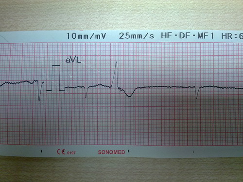
Question: Is it possible to say what the cardiac axis might be just by looking at Lead aVL as shown above?
Answer: It is not possible to say exactly what the cardiac axis is but one can infer in this manner:
1. The current is moving away from aVL. Left axis is the state where the current is moving towards the left side in the segment beyond minus 30 degrees. Therefore lead aVL tells me that this is not left axis.
2. If the current is exactly minus 30 degrees (normal axis) or zero degrees (normal axis), or plus 30 degrees (normal axis), lead aVL should be having a net positive deflection because the electrical scatter is still towards aVL. Therefore the axis is not zero degrees or minus 30 degrees or plus 30 degrees.
3. If the axis is plus 60 degrees, lead aVL should have biphasic deflection (positive and negative deflections are equal) because plus 60 degrees is exactly perpendicular to Lead aVL. Therefore the axis is not plus 60 degrees either.
4. If the axis is plus 90 degrees (normal axis) or beyond 90 degrees (right axis), lead aVL will show a negative deflection because now the electrical scatter is truly away from aVL. Therefore the axis here may be normal (between plus 60 and 90 degrees) or right axis (beyond plus 90 degrees).
Question: Is the ventricular ectopic seen in the given Lead aVL, arising from the right ventricle (RV) or from the left ventricle (LV)?
Answer: If an ectopic beat starts in the RV, it will depolarise the RV first and the LV later. Hence a ventricular ectopic that originates from the RV will have a LBBB pattern. Similarly, a left ventricular ectopic depolarises the LV first and the RV later. Hence it will show a RBBB pattern. So, if a ventricular ectopic shows the LBBB pattern, it is a ventricular ectopic from the RV and if the ectopic beat shows a RBBB pattern, it is a left ventricular ectopic.
The ectopic beat in aVL here (a lead that looks at the left side of the heart) has an LBBB pattern. Therefore it is an ectopic beat that originates from the RV.
Sep 11, 2012
A man who could not feel the left side of his body
A 50 year old man was admitted with a sudden inability to feel the left side of his face, left arm and left lower limb. He could speak and he could move all his limbs. He had been diagnosed to have hypertension a few years earlier but he had discontinued his medicines because he felt well. At the time of admission, his blood pressure was 180/106mm Hg and he was conscious and alert. Neurological examination confirmed that he did not have any deficit other than the hemisensory loss. He began improving about 8 hours after admission and within a day, he had recovered his sensations fully. He was diagnosed as having suffered a transient ischemic attack and treated appropriately.
Discussion
Does the diagnosis of a TIA depend only on how soon a patient recovers form a stroke?
It used to be that a diagnosis of TIA was made when a patient recovers from a cerebrovascular event within 24 hours. Now that we know that some of those with a clinical diagnosis of TIA have a visible infarct on brain imaging, the definition of TIA has changed. A TIA, by the new definition, is a transient cerebrovascular event without any visible lesion on brain imaging.
What about those who recover from a stroke within 24 hours but have a visible infarct on brain imaging?
They should be considered as having suffered a minor stroke.
Can this patient be considered to have suffered a lacunar infarct if his brain imaging shows a visible infarct?
Yes. A pure hemisensory deficit is a manifestation of a lacunar infarct. The various clinical manifestations of a lacunar infarct, and their corresponding lesions, are given below (MedTutor acknowledges, with gratitude, the contribution for this answer by medical student Wong Wai Kit):
Pure motor hemiplegia - the affected artery is the lenticulostriate branch of the middle cerebral artery and the lesion is in the internal capsule.
Pure sensory stroke - the affected artery is thalamogeniculate artery which results in a lesion in the ventral posterio-lateral nucleus of the thalamus.
Clumsy hand syndrome/Dysarthria - the affected artery is the penetrating branch of the basilar artery producing a lesion in dorsal Pons.
Ataxic hemiparesis - the affected artery is the penetrating branch of the basilar artery producing a lesion in the ventral Pons.
Discussion
Does the diagnosis of a TIA depend only on how soon a patient recovers form a stroke?
It used to be that a diagnosis of TIA was made when a patient recovers from a cerebrovascular event within 24 hours. Now that we know that some of those with a clinical diagnosis of TIA have a visible infarct on brain imaging, the definition of TIA has changed. A TIA, by the new definition, is a transient cerebrovascular event without any visible lesion on brain imaging.
What about those who recover from a stroke within 24 hours but have a visible infarct on brain imaging?
They should be considered as having suffered a minor stroke.
Can this patient be considered to have suffered a lacunar infarct if his brain imaging shows a visible infarct?
Yes. A pure hemisensory deficit is a manifestation of a lacunar infarct. The various clinical manifestations of a lacunar infarct, and their corresponding lesions, are given below (MedTutor acknowledges, with gratitude, the contribution for this answer by medical student Wong Wai Kit):
Pure motor hemiplegia - the affected artery is the lenticulostriate branch of the middle cerebral artery and the lesion is in the internal capsule.
Pure sensory stroke - the affected artery is thalamogeniculate artery which results in a lesion in the ventral posterio-lateral nucleus of the thalamus.
Clumsy hand syndrome/Dysarthria - the affected artery is the penetrating branch of the basilar artery producing a lesion in dorsal Pons.
Ataxic hemiparesis - the affected artery is the penetrating branch of the basilar artery producing a lesion in the ventral Pons.
Subscribe to:
Posts (Atom)